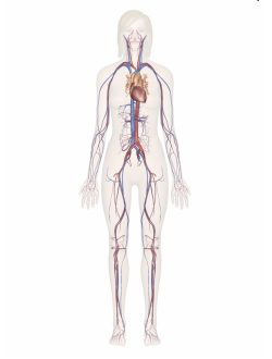
Your pulse comes from the heart: each contraction sends a pressure wave through the arteries. Veins have low, steady pressure, so there's no pulse there. Blood pressure is usually taken at the brachial artery.
Major veins
Subclavian: under the collarbone
Superior vena cava: upper chest, from the upper body
Inferior vena cava: abdomen, from the lower body
Great saphenous: inner leg, foot to groin
The Heart
Circulatory System Study Sheet 3 of 10 Chambers, valves and layers
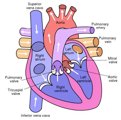
Right side · oxygen-poor
Superior & inferior vena cava
Bring deoxygenated blood from the body
Right atrium
Receives blood from the vena cavae
Tricuspid valve
Between right atrium and right ventricle; prevents backflow
Right ventricle
Pumps blood to the lungs
Pulmonary semilunar valve
Between right ventricle and pulmonary artery
Apex
The bottom tip. The heartbeat is heard best here with a stethoscope.
Interventricular septum
The wall dividing the right and left ventricles.
The coronary arteries branch off the aorta and supply the heart muscle itself. The left side is stronger because it pumps blood to the whole body.
Left side · oxygen-rich
Pulmonary veins
Bring oxygenated blood from the lungs
Left atrium
Receives blood from the pulmonary veins
Bicuspid / mitral valve
Between left atrium and left ventricle; prevents backflow
Left ventricle
Pumps blood to the whole body; thickest wall
Aortic valve
Between left ventricle and aorta
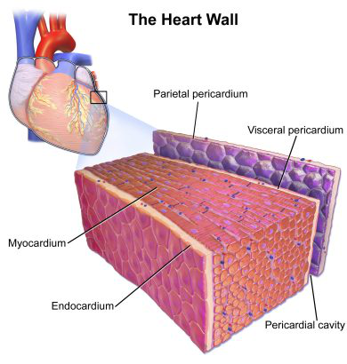
Outer layer
Pericardium
Inflammation of this layer is called pericarditis.
Middle layer
Myocardium
The muscle layer responsible for the heart's contraction.
Inner layer
Endocardium
Smooth lining made of endothelial cells.
The Path of Blood Through the Heart and Body
Circulatory System Study Sheet 4 of 10 Follow the numbers 1 → 15
Step by step
1Deoxygenated blood leaves the body tissue
2Superior & inferior vena cavabring it back to the heart
3Right atrium
4Tricuspid valve
5Right ventriclepumps blood to the lungs
6Pulmonary valve
7Pulmonary artery
8Both lungsgas exchange in the alveoli
9Pulmonary veins
10Left atrium
11Bicuspid (mitral) valve
12Left ventriclepumps blood to the whole body
13Aortic valve
14Aorta
15Oxygenated blood reaches the body cells
Remember
Arteries carry blood away from the heart. Veins carry blood toward it.
The exception: the pulmonary artery carries oxygen-poor blood, and the pulmonary veins carry oxygen-rich blood.
The right side pumps to the lungs; the left side pumps to the whole body, so the left ventricle is strongest.
"TRI before you BI": blood passes the tricuspid valve first, the bicuspid second.
The Electrical System of the Heart
Circulatory System Study Sheet 5 of 10 How a heartbeat starts and spreads
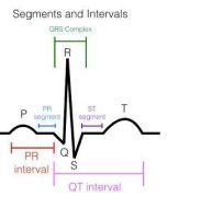
T wave = ventricles relax (ventricular repolarization)
The electrocardiograph (ECG/EKG) records the heart's electrical activity.
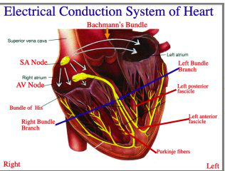
The signal's path
1
SA (sinoatrial) node — the PACEMAKER Top of the right atrium. Its signal spreads over both atria, making them contract.
↓
2
AV (atrioventricular) node Between the right atrium and right ventricle. Passes the signal from the atria to the ventricles.
↓
3
Bundle of His Carries the signal down between the ventricles.
↓
4
Right and left bundle branches Split the signal toward each ventricle.
↓
5
Purkinje fibers Deliver the signal through the ventricles, causing ventricular contraction.
Sends blood to lungs & body
The Bundle of His, bundle branches and Purkinje fibers, because they trigger the ventricles.
When the system fails
One treatment is an artificial pacemaker.
Blood Pressure
Circulatory System Study Sheet 6 of 10 Measured in millimeters of mercury (mmHg)
120
80
mmHg
Usually measured at the brachial artery in the upper arm.
Top number
Systolic pressure
Pressure of blood on the arteries during ventricular contraction (systole).
Bottom number
Diastolic pressure
Pressure on the arteries during ventricular relaxation (diastole).
Hypertension
Called "the silent killer." It can lead to an aneurysm or a hemorrhagic stroke (CVA).
What changes blood pressure?
Highly conditioned aerobic athletes: often lower, because a stronger heart pumps more blood with each beat.
Plaque buildup in the arteries: often higher, because narrowed, stiffer arteries resist flow.
Common blood pressure medications
ACE inhibitors (lisinopril)
Beta blockers (metoprolol)
Diuretics (hydrochlorothiazide)
Calcium channel blockers (amlodipine)
Blood
Circulatory System Study Sheet 7 of 10 What blood is made of
Plasma on top · thin layer of leukocytes & thrombocytes · erythrocytes on the bottom
10–12 pints
in the average adult, about 7–8% of body weight. Blood is filtered by the kidneys.
Plasma · about 55%
The yellow liquid part of blood, made mostly of water.
Plasma proteins
Fibrinogen: needed for clotting; made in the liver
Albumin: made in the liver; maintains osmotic pressure and blood volume; the most abundant plasma protein
Prothrombin: helps blood coagulate; needs vitamin K (measured by the PT or "Protime" test)
Erythrocytes and leukocytes are full cells. Thrombocytes are only cell fragments.
Erythrocytes · red blood cells · about 45%
Made in the long bones by hematopoiesis. They transport O2 and CO2 using the protein hemoglobin. Shape: a biconcave disc.
Low hemoglobin can be a sign of anemia. Normal: men about 13.5–17.5 g/dL, women about 12.0–15.5 g/dL.
Leukocytes · white blood cells
Protect against and fight pathogens.
Type
Job
Lymphocytes
Create antibodies; protect against cancer
Monocytes
Phagocytosis of debris and foreign particles
Granulocytes
Neutrophils
First responders during an infection
Eosinophils
Increase during parasitic infection and malaria
Basophils
Release heparin and histamine to promote inflammation
Thrombocytes · platelets
Fragments of megakaryocytes. Necessary for blood clotting; removed after about 10 days.
Blood Types and Rh Factor
Circulatory System Study Sheet 8 of 10 Antigens, antibodies and who can donate to whom
ABO blood types
Blood type is set by an antigen, a protein marker on the surface of red blood cells. The body makes antibodies against antigens it doesn't have.
Type
Antigen
Antibodies
Donates to
Receives from
A
Anti-B
A, AB
A, O
B
Anti-A
B, AB
B, O
AB
None
AB
All types
O
Anti-A and anti-B
All types
O
Why? O cells have neither antigen, so both A and B look foreign. AB cells have both, so neither looks foreign.
Erythroblastosis fetalis
A concern when the mother is Rh− and the father is Rh+. If the baby is Rh+, the mother builds antibodies; in a later Rh+ pregnancy they attack the baby's red blood cells and can cause a miscarriage. Prevented with RhoGAM.
Transfusion reaction
Happens when a person receives an incompatible blood type. The immune system attacks the incompatible blood.
Rh factor
The rhesus (Rh) factor is another protein that is either present or absent on red blood cells. Has it: positive. Lacks it: negative.
Most common: O+ then A+
Who can give blood to whom
Receives ↓ / Gives →
O-
O+
A-
A+
B-
B+
AB-
AB+
O-
O+
A-
A+
B-
B+
AB-
AB+
Negative types can donate to positive and negative, but receive only negative. Positive types can receive both, but donate only to positive.
O−
Universal donor
AB+
Universal recipient
Disorders of the Blood and Blood Vessels
Circulatory System Study Sheet 9 of 10 Grouped by type: blockage, hemorrhage, other
Blockage
Blood can't get through
Coronary artery diseaseCoronary arteriesPlaque narrows the arteries to the heart muscle (atherosclerosis). Symptom: angina (chest pain). Increase exercise, quit smoking, reduce cholesterol.
Thrombus / embolismAny artery or veinA thrombus is a clot. An embolism is a clot that breaks off and travels.
Pulmonary embolismPulmonary arteriesA clot that travels to the lungs. A medical emergency!
Ischemic stroke (CVA)Carotid and cerebral arteriesFace droop, arm numbness, slurred speech. Clot-busting (thrombolytic) medication; best within 4 hours.
Peripheral artery diseaseLeg arteries (femoral, popliteal)Leg pain that improves with rest; discolored legs/toes. Reduce cholesterol, surgery. Untreated: gangrene.
Hemorrhage
The vessel wall fails and bleeds
An aneurysm is a ballooning of an artery with a thinning wall, caused by a weakened blood vessel.
Brain aneurysmCerebral arteriesSame symptoms as a stroke: weakness on one side, slurred speech, severe headache, confusion. Immediate care; surgical repair.
Hemorrhagic stroke (CVA)Cerebral arteriesA brain aneurysm ruptures and bleeds. Surgery to stop the bleeding.
Aortic aneurysmAortaMay have no symptoms, or back or abdominal pain. Monitoring or surgical repair; a rupture causes immediate death.
Other
Pressure and pooling
HypertensionAll arteriesExcessive pressure on the artery walls. Can lead to an aneurysm or a hemorrhagic stroke.
AnginaCoronary arteries (narrowed)Chest pain; can be caused by coronary artery disease.
Varicose veinsLeg veins (great saphenous)Slow blood flow stretches the valves; pooled blood makes the vein walls bulge. Causes: obesity, prolonged standing, age, pregnancy. Compression hose, elevate legs, surgery/ablation.
Disorders of bloodAnemia: RBCs can't carry enough O2. Many causes.Sickle cell anemia: genetic; misshapen RBCs bind less O2.Hemophilia: genetic bleeding disorder; blood doesn't clot properly.Leukemia: a type of blood cancer.
Heart Disorders
Circulatory System Study Sheet 10 of 10 Rhythm, electrical, plumbing, pumping and valve problems
Rhythm
Arrhythmia
Bradycardia: heart beats too slowly.
Tachycardia: heart beats too fast.
Dysrhythmia: an abnormal rhythm.
60–100
normal heart rate
Electrical
Cardiac arrest
The heart suddenly stops. Causes: ventricular fibrillation, severe heart attack, a genetic defect or another electrical problem.
Symptoms: sudden collapse, no pulse, no breathing.
Treatment: 911, CPR.
Plumbing
Myocardial infarction
A heart attack, caused by coronary artery disease. Heart muscle cells die.
Symptoms: angina, jaw or shoulder pain, nausea, sweating, fainting, fluid in the lungs.
Treatment: 911 or the ED, blood thinners, bypass surgery.
Pumping
Heart failure
Also called congestive heart failure. The ventricles can't contract effectively and blood pools.
Left side fails: fluid in the lungs, shortness of breath. Right side fails: swelling in the body tissues.
Treat the cause. There is no cure.
Valves
Heart murmur
A defect in the valves: blood leaks backward, or a valve opening is narrowed.
Treatment: usually monitoring.
Heart attack vs cardiac arrest vs heart failure
Heart attack = a plumbing problem. A blocked coronary artery cuts off oxygen and part of the heart muscle dies.
Cardiac arrest = an electrical problem. The heart suddenly stops pumping.
Heart failure = a pumping problem. The ventricles become too weak to pump well.
How they relate: a heart attack can trigger cardiac arrest right away, and the damage it leaves can weaken the heart over time, leading to heart failure.